Age-related macular degeneration
- What is age-related macular degeneration (AMD)?
- What are the different types of age-related macular degeneration?
- What are the symptoms of macular degeneration?
- What causes age-related macular degeneration?
- What are the risk factors for age-related macular degeneration?
- Can macular degeneration cause complications?
- How is macular degeneration diagnosed?
- What are the treatments for macular degeneration?
- Can macular degeneration be prevented?
- Living with macular degeneration
- Featured age-related macular degeneration articles
Introduction
There are more than 196 million people worldwide with macular degeneration, according to the World Health Organization (WHO). In the United States, nearly 13 percent of adults older than 40—or almost 20 million people—have the eye disease, reports the Centers for Disease Control and Prevention (CDC), making it the most common cause of vision loss among older adults. But macular degeneration it's an inevitable part of aging and there are ways to reduce your risk of developing it.
Learn more about the different types of macular degeneration, symptoms of the condition, as well as causes and treatment options. Discover tips for protecting your vision and find out how to live with macular degeneration while maintaining your sense of well-being.
What is age-related macular degeneration (AMD)?
Macular degeneration is an eye disorder that affects your macula, the part of the eye responsible for straight-ahead vision. The macula is part of the retina, the tissue located at the back of the eye that is sensitive to light. Your macula is made up of specialized nerve cells called photoreceptors that convert light into nerve impulses. Photoreceptors come in two shapes: rods and cones. Rods control vision in low-light situations while cones help your eyes see color and adapt to changes in light.
Macular degeneration also affects the central portion of the macula within the retina called the fovea. Your fovea controls your ability to discern shapes and details from a distance, also known as your visual acuity.
Macular degeneration impedes your ability to see these fine details from up close and further away. While the condition affects the center of your field of vision, your peripheral (side) vision stays intact. As the name age-related macular degeneration implies, this condition progressively blurs your visual acuity as you age.
Juvenile macular degeneration
Macular degeneration can also affect children and young adults. The rare eye disorders that affect this age group are known collectively as juvenile macular degeneration (JMD), also called juvenile macular dystrophy.
Like age-related macular degeneration, JMD also causes central vision loss. The most common type of JMD is Stargardt disease, followed by Best’s disease (also called Best’s vitelliform retinal dystrophy), and juvenile retinoschisis. But unlike age-related macular degeneration, which develops as part of the aging process, juvenile macular degeneration is inherited from parents.
What are the different types of age-related macular degeneration?

There are two main types of age-related macular degeneration:
Dry macular degeneration
Macular degeneration almost always starts off as dry macular degeneration. Around 80 to 90 percent of people with the condition have the dry type, according to a 2022 review of studies published in Frontiers in Neuroscience.
As the retina deteriorates, small yellow waste deposits called drusen form under the macula. The accumulation of drusen causes your macula to grow thin, dry, and inflamed, leading to the breakdown of its light-sensitive cells and underlying supportive tissue.
Sudden blurry vision and other symptoms can occur in one or both eyes, although it usually affects both eyes. When only one eye is affected or one eye has more damage than the other, you may not notice much change to your vision. That’s because your unaffected or less affected eye may compensate for deterioration in the other eye.
Dry macular degeneration is also known as atrophic macular degeneration. This refers to the atrophy (wasting away) of a layer of cells in the retina called the retinal pigment epithelium (RPE). These cells play an important role in the eye’s ability to absorb light.
Dry macular degeneration tends to progress slowly, advancing in stages from early to intermediate to the advanced stage of dry macular degeneration called geographic atrophy. This late-stage macular degeneration causes blind, blank, or dark spots in your visual field. It’s known as “geographic” atrophy because the atrophied and dead cells merge to resemble features of a map when viewed on an eye exam.
Wet macular degeneration
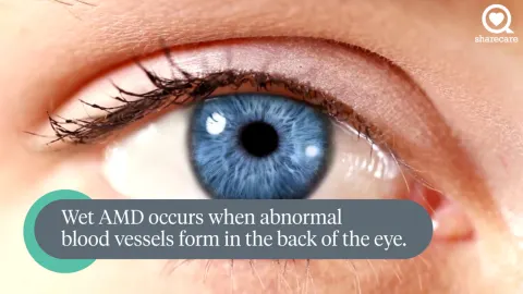
Dry macular degeneration can progress to wet macular degeneration at any point. This advanced form of the eye disease is also called neovascular macular degeneration (NMD). The word neovascular refers to the formation of new blood vessels, which emerge in a layer of the eye just behind the retina called the choroid.
During this phase of the disease, known as choroidal neovascularization (CNV), new blood vessels can bleed or leak fluid, which causes your macula to bulge or lift from its usual flat-lying position. NMD can thus distort and destroy your central vision more rapidly and aggressively than the typically slower progression of dry macular degeneration.
What are the symptoms of macular degeneration?
Few, if any, symptoms occur during the early stages of the disease. But an HCP may be able to spot early signs of macular degeneration during a routine eye exam and to distinguish it from other eye diseases that cause vision loss, such as cataracts or glaucoma.
For this reason, it’s important that everyone should receive regular routine eye exams as part of an overall wellness plan. The HCPs you may see for these exams include ophthalmologists (medical doctors who specialize in eye care) or optometrists (licensed eye specialists who provide primary vision care).
Early warning signs your HCP may detect during a comprehensive vision exam include drusen on the surface of your retina, as well as changes to the pigment color of your macula. Early symptoms of macular degeneration might also include mild changes to your reading capacity, especially when lights are dim. You could also experience sudden blurry vision or problems seeing faces clearly, or you may have no signs or symptoms.
If the disease progresses past its early stage, symptoms can include:
- Blurry vision that worsens or blank spots in your central field of vision that grow larger and darker
- Colors that seem less vibrant
- Needing more light when you’re reading or viewing things up close or having a hard time adjusting to low light levels
- Printed words that look increasingly blurry when you try to read them
- Reduced central vision in one or both eyes
- Visual distortions (for example, straight lines that look wavy or objects that appear smaller than they really are)
What causes age-related macular degeneration?
Age-related macular degeneration occurs as a result of the deterioration of various structures that support the part of the retina called the macula. Beneath the macula is the choroid, a layer of tissue that delivers blood to the macula. Atop the macula is a layer of retinal pigment epithelium (RPE) cells that transport nutrients from the choroid to the macula and move macular waste products out via the choroid.
As you age, your RPE becomes thinner, which affects its ability to move nutrients and waste. As this happens, macular damage and vision impairment may occur due to the buildup of waste products and the lack of blood and nutrients.
The RPE also nourishes a layer of tissue called Bruch’s membrane that helps deliver nutrients and oxygen to the retina, while also removing waste products. It’s normal for Bruch’s membrane to thicken as you age, but age-related macular degeneration can cause it to grow excessively thick. This further hampers nutrient, oxygen, and waste exchange between the RPE and choroid, which impairs your ability to see colors and images clearly, especially in low-light situations. In wet AMD, new blood vessels from the choroid can grow through breaks in Bruch's membrane into the RPE, causing bleeding below the retina.
What are the risk factors for age-related macular degeneration?
Along with being older, risk factors for age-related macular degeneration include:
- Being white or Hispanic
- Being far-sighted
- Cardiovascular and metabolic diseases such as high blood pressure and atherosclerosis
- Diabetic retinopathy, elevated high-density lipoprotein (aka HDL or “good” cholesterol), high systolic blood pressure (the top number in your blood pressure reading), and obesity, especially if you have diabetes
- Diets high in saturated fats
- Elevated C-reactive protein, a marker that signals inflammation in the body
- Family history of macular degeneration among close biological relatives, such as a parent and sibling
- Inherited gene variants, also called gene mutations, such as those involving complement factor H (CFH) and human high temperature requirement serine protease A1 (HTRA1)
- Light-colored irises (for example, having blue eyes)
- Smoking, which raises risk and hastens disease progression
- Ultraviolet (UV) light exposure from the sun and other UV sources such as fluorescent lighting and video display terminals
- Blue light exposure from smart phones, computers, and other electronic devices with screens
Can macular degeneration cause complications?
Macular degeneration can make it harder for you to carry out certain visual tasks such as cooking, driving, repairing household items, and reading. These visual changes can impact your lifestyle, activities, and social life, which can lead to depression and anxiety.
The eye disease can also raise the risk of:
- Falls and injuries: Central vision loss can increase your chances of falling and getting hurt, especially if you already have a higher risk due to health conditions that affect your balance or medicines you take such as opioids, muscle relaxants, sedatives, and blood pressure medicine.
- Blindness: Severe macular degeneration can cause you to become legally blind, defined as having visual acuity of 20/200 (one-tenth of 20/20 vision) or worse in your best-seeing eye while wearing corrective lenses such as glasses.
- Retinal detachment: This is when tissues in your retina separate from choroid blood vessels, which can cause vision loss.
- Visual hallucinations: Charles Bonnet syndrome, a condition associated with vision loss in which you see things that aren’t there, is more likely to occur during late-stage macular degeneration.
How is macular degeneration diagnosed?

Your primary care provider can look for changes in your eyes during a dilated eye exam. After placing drops in your eyes to dilate (widen) your pupils, your provider may use a handheld device equipped with a light source, mirrors, and lenses called an ophthalmoscope to examine the back of your eyes. If they detect changes that are concerning or indicative of age-related macular degeneration, they’ll likely refer you to an eye specialist such as an optometrist or ophthalmologist.
An eye specialist will likely use a dilated eye test called a slit lamp exam to more closely examine the interior structures of your eyes. During the exam, you’ll sit in a chair facing the device with your chin and forehead resting against a brace to keep your head steady. On the other side of the slit lamp, the eye specialist will shine a bright light into each eye and examine your eyes through a microscope. They will look for blood, fluid, drusen deposits, or a blotchy or mottled appearance caused by changes to your macular pigment.
After the exam, you’ll need someone to take you home because you won’t be able to drive. Your eyes will be temporarily blurry and sensitive to light as a result of dilation.
During your visit, your eye provider may also ask you to read a Snellen eye chart from 20 feet away. You may recognize this chart by the large “E” at the top of it. They may also use an Amsler grid to test your visual acuity, after which they’ll ask you to take the grid home to monitor for vision changes that might occur.
Self-testing with the Amsler grid
The Amsler grid is a simple square with a grid pattern and a dot in the middle. When used properly, the design can spot blurry or blank spots in your central field of vision.
If you show early signs of age-related macular degeneration or have risk factors for the disease, the American Academy of Ophthalmology (AAO) recommends testing your visual acuity daily with an Amsler grid. Follow these steps to perform the test:
- Wear your reading glasses, if you use them, and hold the grid 12 to 15 inches away from your eyes in good light.
- Cover one eye and look directly at the dot in the center of the grid with your uncovered eye.
- Keep your eye focused on the dot and note whether the grid lines appear straight or if any lines or areas of the grid look blurry, bent, wavy, dim, or blank.
- Test your other eye following the same sequence.
Let your optometrist or ophthalmologist know right away if any lines or areas of the grid look blurry, curved, darkened, or distorted.
Advanced testing for macular degeneration
Your HCP may order one or more tests to confirm a macular degeneration diagnosis. These advanced tests involve looking at images of the inside, back surface of your eye, which consists of your macula, retina, optic disc, fovea, and blood vessels and is known collectively as the fundus.
These tests include:
- Fundus fluorescein angiography (FFA): The FFA involves injecting a yellow fluorescent dye called fluorescein into a vein and photographing the flow of blood. The presence of fluorescent patches points to leakage in the retina and choroid.
- Indocyanine green angiography: This test also involves injecting dye into the bloodstream. It’s used to confirm FFA findings or to identify blood vessel problems deeper in the retina.
- Fundus autofluorescence imaging (AF): This noninvasive imaging method uses natural fluorescent structures in the body called fluorophores to assess the retina. These fluorophores, which include RPE cells, light up when exposed to various wavelengths of light. AF is often used to test for late dry macular degeneration or geographic atrophy since atrophied areas of the eye don’t light up.
- Optical coherence tomography (OCT): This noninvasive test uses infrared light waves to create detailed, cross-section images of the retina and choroid layer. OCT can spot drusen, new blood vessels, blood or fluid leakage, and retinal thinning, thickening, and swelling.
- OCT angiography: Although still used mainly as a research tool, this noninvasive test is being used more often to look for abnormal blood vessel growth in the macula.
What are the treatments for macular degeneration?
While there is no cure for the disease, there are a variety of treatment and management strategies for macular degeneration that can help control symptoms and slow progression. Your HCP will discuss options with you based on the type and stage of your disease.
These include:
Nutrition supplements for dry macular degeneration
If you have a lot of drusen, taking a specific combination of nutrition supplements for eye health might help curb the progression of dry macular degeneration. But bear in mind that these supplements aren’t likely to help with advanced age-related macular degeneration.
Supplement recommendations are based on the most recent Age-Related Eye Disease Study (AREDS), called AREDS2, from the National Eye Institute (NEI).
Specially formulated AREDS2 supplements include:
- Copper (2 milligrams)
- Lutein (10 milligrams)
- Vitamin C (500 milligrams)
- Vitamin E (400 international units)
- Zeaxanthin (2 milligrams)
- Zinc (80 milligrams)
AREDS2 supplements can be bought over the counter but before you add them to your macular degeneration treatment plan, talk with your HCP about their benefits and risks and how they might interact with other medicines and supplements you take.
Injectable medicine to treat dry macular degeneration
In February 2023, the U.S. Food and Drug Administration (FDA) approved the use of pegcetacoplan to treat geographic atrophy (advanced dry macular degeneration). It was the first such medicine or therapy approved to treat this form of macular degeneration and involves injection of the medicine into your eye through a procedure called intravitreal injection (IVI). Pegcetacoplan helps prevent retinal tissue atrophy and has been shown to slow the growth of geographic atrophy lesions.
The FDA approved another medicine, avacincaptad pegol, for treatment of geographic atrophy in August 2023. It also involves an injection into the eye. This treatment addresses the source of retinal cell death and has been shown to significantly slow the progression of geographic atrophy after one year of use.
Stem cell therapy for dry macular degeneration
Stem cell therapy hasn’t been approved as a treatment for macular degeneration, although numerous clinical trials are underway. In general, studies looking at transplantation techniques show promise that human stem cells may be able to repair retinal damage caused by the disease.
A 2022 review and analysis of randomized clinical trials published in Stem Cell Research & Therapy found that stem cell therapy may help improve best-corrected visual acuity (BCVA) in people with dry macular degeneration. BCVA measures your visual acuity while wearing glasses or contact lenses.
Clinical trials have focused mainly on stem cell therapy for dry macular degeneration. The approach aims to replace damaged RPE cells with healthy ones. This may help protect and preserve the health and function of remaining photoreceptors and repair damage to these light-sensitive cells.
Anti-VEGF medications to treat wet macular degeneration
Anti-VEGF therapy prevents a protein called endothelial growth factor (VEGF) from forming new blood vessels in your macula. Anti-VEGF medicines are given through IVI. They’re often the first treatment your HCP will recommend for wet macular degeneration.
Anti-VEGF medicines include:
- Aflibercept
- Bevacizumab
- Brolucizumab
- Faricimab-svoa
- Ranibizumab
Anti-VEGF medicines help shrink blood vessels and absorb fluid under your retina, which may help you recover some of the vision you lost to macular degeneration. To maintain their therapeutic effects, these IVIs must be given every four to six weeks.
Other treatment options for wet macular degeneration include:
Photodynamic therapy to treat wet macular degeneration
During photodynamic therapy, your HCP injects the medicine verteporfin into a vein in your arm. Once the medicine travels through your bloodstream to blood vessels in your macula, your HCP shines a focused beam of light from a special laser onto the vessels to activate the medicine. As a result, the blood vessels close and stop leaking, which may help improve your vision and slow down the rate of vision loss.
You’ll likely need multiple sessions of photodynamic therapy because treated vessels may reopen. While photodynamic therapy was once the standard treatment for wet macular degeneration, anti-VEGF therapy is now used more frequently.
Laser photocoagulation for wet macular degeneration
During laser photocoagulation, your HCP will use a high-energy laser beam to seal leaky blood vessels under your macula. Multiple sessions are needed since new abnormal blood vessels can form.
Laser photocoagulation can produce blind spots due to scarring. It can’t treat damaged blood vessels under the center of the macula. And it’s also less likely to be successful if your macula has a lot of damage. This is another older procedure that is no longer commonly used.
Can macular degeneration be prevented?
There’s no certain way to prevent the disease but lifestyle changes can help you manage macular degeneration and lower your risk for it. Aim to:
- Be physically active on a regular basis.
- Maintain a healthy weight and healthy blood pressure and cholesterol levels.
- Quit smoking or never start.
- Wear a hat with a wide brim and sunglasses with maximum UV protection when you’re outdoors. The maximum level of UV protection for sunglasses is UV 400, which blocks 99.9 percent of UVA and UVB rays.
- Maintain routine visits for eye care.
Age-related macular degeneration diet
Eating a nourishing diet for macular degeneration can help preserve your eye health and lower your risk for macular degeneration. A 2022 review of studies published in Acta Opthalmologica found that in addition to taking AREDS2 supplements, a wholesome diet that emphasizes nutrients beneficial for eye health can lower the chances of getting early macular degeneration or the likelihood that the disease will progress if you already have it.
These nutrients include:
- Beta carotene: found in green, yellow, and orange fruits and vegetables such as broccoli, carrots, cantaloupe, spinach, sweet potatoes, tomatoes, and winter squash
- Calcium: found in almonds, sardines with bones, dairy products such as cow’s milk and cheese, and leafy greens such as Bok choy, collard greens, kale, mustard greens, and turnip greens
- Copper: found in beef liver, lobster, oysters, shiitake mushrooms, cashews, and sesame seeds
- Folate: found in beef liver, eggs, black-eyed peas, asparagus, Brussel sprouts, spinach, and other dark leafy greens
- Lutein and zeaxanthin: found in egg yolks, dark green vegetables such as kale, parsley, spinach, broccoli and peas, as well as colorful fruits such as honeydew melon, kiwis, and grapes
- Lycopene: found in red, orange, and pink fruits such as apricots, papaya, pink guavas, pink grapefruits, and watermelon
- Magnesium: found in pumpkin seeds, chia seeds, almonds, spinach, cashews, peanuts, black beans, edamame, peanut butter, and brown rice
- Niacin, also known as vitamin B3: found in liver, chicken breast, turkey, tuna, anchovies, beef, pork, peanuts, mushrooms, green peas, white potatoes, and whole wheat bread and pasta
- Vitamin A: found in beef liver, lamb liver, King mackerel, salmon, eggs, bluefin tuna, trout, oysters, clams, goat cheese, cheddar cheese, and whole cow’s milk
- Vitamin B6: found in avocado, chickpeas, bananas, green peas, sweet potatoes, spinach, carrots, beef, chicken liver, salmon, yellowfin and albacore tuna, cow’s milk, and ricotta cheese
- Vitamin C: found in guava, kiwi, sweet red pepper, oranges, strawberries, papaya, pineapple, mango, broccoli, cauliflower, and Brussels sprouts
- Omega-3 fatty acids, specifically eicosapentaenoic acid (EPA) and docosahexaenoic acid (DHA): found in shellfish such as oysters and cold-water fatty fish such as salmon, mackerel, tuna, herring, and sardines
Following the Mediterranean diet may also lower your risk for developing early macular degeneration and may slow down the progression to the advanced stages of the disease. The diet focuses on consuming:
- Plant-based oils rich in poly- and monounsaturated fats like olive oil instead of saturated fats such as butter
- Whole plant foods such as fruits, vegetables, legumes, whole grains, and nuts
- Moderate amounts of fish, poultry, dairy, and red wine
- Limited amounts of red meat
When to see your healthcare provider
Be sure to contact your HCP if you notice any macular degeneration symptoms, especially if these symptoms are new, getting worse, or persisting. If you experience severe symptoms, be sure to get treatment right away.
Severe symptoms might include:
- Darkening or shadows covering part of your vision
- Fleeting flashes of light
- Painful eye inflammation and sensitivity to light
- Pressure behind your eye
- Sudden or significant increase in eye floaters such as dark spots, flecks, squiggly lines, and threads that temporarily dart across your field of vision
Living with macular degeneration

Living with macular degeneration means making the most of your vision. You can still do many of your favorite things using tools to improve low vision, such as magnifiers, audio books, and smart devices that allow you to adjust print and image size.
You can also ask your HCP to refer you for low-vision rehabilitation. You’ll work with an occupational therapist or other trained specialist to learn ways to adapt to your changing vision while maintaining your independence.
This might include helping you access support services and teaching you how to use low-vision tools, making use of the part of your vision that’s intact such as your peripheral vision, and making modifications to your home such as:
- Applying large-print stickers or tape on your thermostat, oven, and refrigerator
- Installing handrails on your stairs and steps, grab bars in your bathroom, and extra lights (such as nightlights and overhead, task, and stair lights)
- Labeling your medicines with large-print stickers or tape
- Marking light switches, electrical outlets, and the edges of steps with bright tape
- Placing raised labels on your computer keys
- Keeping walkways open and clear, removing area rugs, and making thresholds flush with your floors and carpet to eliminate tripping hazards
The vision loss caused by macular degeneration can impact your mental health along with your physical health. It can leave you feeling isolated, alone, and frustrated.
Remember that you’re not alone and help is available. Talk to your HCP about whether it would make sense for you to speak with a licensed mental health provider. Expressing your emotions and frustrations and problem-solving in a safe, supportive environment can help you maintain your sense of well-being.
They can also help you develop coping skills for the stressors and challenges that come with having the disease and connect you with in-person and online support groups to find common cause with other patients.
In the meantime, it’s important to keep up a regular schedule of follow-up visits with your primary eye care provider to make sure your macular degeneration management plan stays on track.
Featured age-related macular degeneration articles
Alcalde I, Sánchez-Fernández C, Martín C, et al. Human stem cell transplantation for retinal degenerative diseases: Where are we now? Medicina. 2022;58(1):102.
American Macular Degeneration Foundation. Diagnosing Age-related Macular Degeneration. Accessed March 21, 2023.
American Macular Degeneration Foundation. Dry Macular Degeneration. Accessed March 20, 2023.
American Macular Degeneration Foundation. Macular Degeneration Symptoms. Accessed March 20, 2023.
American Macular Degeneration Foundation. Photograph of the Retina Showing Macula With Drusen. Accessed March 20, 2023.
American Macular Degeneration Foundation. Risk Factors for Macular Degeneration. Accessed March 20, 2023.
American Macular Degeneration Foundation. Wet Macular Degeneration. Accessed March 20, 2023.
American Macular Degeneration Foundation. What Is Macular Degeneration? Accessed March 20, 2023.
Bonavitacola J. FDA Approves New Treatment for Geographic Atrophy. The American Journal of Managed Care. Published August 7, 2023.
Boyd B. What Is Macular Degeneration? American Academy of Ophthalmology. Published February 10, 2022.
Boyd K. Have AMD? Save Your Sight With an Amsler Grid. American Academy of Ophthalmology. Published May 26, 2020.
Csader S, Korhonen S, Kaarniranta K, Schwab U. The effect of dietary supplementations on delaying the progression of age-related macular degeneration: A systematic review and meta-analysis. Nutrients. 2022;14(20):4273.
Centers for Disease Control and Prevention. Learn About Age-Related Macular Degeneration. Last reviewed November 23, 2020.
Centers for Disease Control and Prevention. Prevalence of Age-Related Macular Degeneration (AMD). Last reviewed October 31, 2022.
Chambers WA. NDA 217171: NDA Approval. United States Food and Drug Administration. Issued February 17, 2023.
Cho YK, Park DH, Jeon IC. Medication trends for age-related macular degeneration. Int J Mol Sci. 2021;22(21):11837.
Deng Y, Qiao L, Du M, et al. Age-related macular degeneration: Epidemiology, genetics, pathophysiology, diagnosis, and targeted therapy. Genes Dis. 2021;9(1):62-79.
ElSheikh RH, Chauhan MZ, Sallam AB. Current and novel therapeutic approaches for treatment of neovascular age-related macular degeneration. Biomolecules. 2022;12(11):1629.
Leng T, Schwartz J, Nimke D, et al. Dry age-related macular degeneration: Distribution of visual acuity and progression risk in a large registry. Ophthalmol Ther. 2023;12(1):325-340.
Li L, Yu Y, Lin S, Hu J. Changes in best-corrected visual acuity in patients with dry age-related macular degeneration after stem cell transplantation: Systematic review and meta-analysis. Stem Cell Res Ther. 2022;13(1):237.
Mayo Clinic. Dry Macular Degeneration. Last updated November 23, 2022.
Mayo Clinic. Wet Macular Degeneration. Last updated February 21, 2023.
Mount Sinai. Macular Degeneration. Accessed March 20, 2023.
National Eye Institute. Age-Related Macular Degeneration. (AMD). Last updated June 22, 2021.
Pameijer EM, Heus P, Damen JAA, et al. What did we learn in 35 years of research on nutrition and supplements for age-related macular degeneration: A systematic review. Acta Ophthalmol. 2022;100(8):e1541-e1552.
Porter D. Diet and Nutrition. American Academy of Ophthalmology. Last updated November 2, 2020.
Posvar W, Pucker A. A Deeper Look at Pegcetacoplan. Optometry Times. Published October 16, 2023.
Rubner R, Li KV, Canto-Soler MV. Progress of clinical therapies for dry age-related macular degeneration. Int J Ophthalmol. 2022;15(1):157-166.
Ruia S, Kaufman EJ. Macular Degeneration. StatPearls [Internet]. Last updated August 3, 2022
Turbert D. What Is Juvenile Macular Dystrophy? American Academy of Ophthalmology. Published November 8, 2022.
United States Food and Drug Administration. NDA 217171: Highlights and Full Prescribing Information. Last updated February 2023.
Vyawahare H, Shinde P. Age-related macular degeneration: Epidemiology, pathophysiology, diagnosis, and treatment. Cureus. 2022;14(9):e29583.
Wang Y, Zhong Y, Zhang L, Wu Q, Tham Y, Rim T, H, Kithinji D, M, Wu J, Cheng C, Liang H, Yu H, Yang X, Liu L. Global incidence, progression, and risk factors of age-related macular degeneration and projection of disease statistics in 30 years: A modeling study. Gerontology 2022;68:721-735.
Wong JHC, Ma JYW, Jobling AI, et al. Exploring the pathogenesis of age-related macular degeneration: A review of the interplay between retinal pigment epithelium dysfunction and the innate immune system. Front Neurosci. 2022;16:1009599.
World Health Organization. World Report on Vision. Last updated 2019.
Porter D. What is a Slit Lamp? American Academy of Ophthalmology. Published April 23, 2018.
Porter D. What Is Charles Bonnet Syndrome? American Academy of Ophthalmology. Published September 21, 2022.


